Research
Study of Ultrafast Kinetics
Interrogation of nuclear dynamics at the temporal resolution of nuclear motion
Ultrafast Raman Loss Spectroscopy
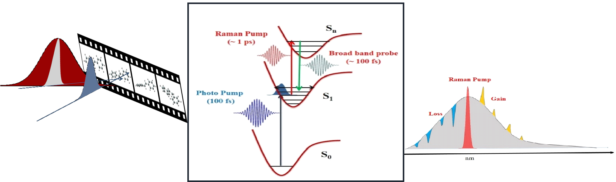
Ultrafast stimulated Raman has provided a new way to acquire vibrational signature of the transient species in a photochemical transformation. Using ultrashort pulses (100 femtoseconds) one can create and monitor transient structural change through nonlinear stimulated Raman technique. We have developed a new method to acquire vibrational Raman signature free from fluorescence background. Combining three optical pulses namely Photo-pump (100 fs), broadband probe (100 fs) and Raman pump (1 ps) and with sophisticated data acquiring method we could observe Raman signature of the short-lived species. By applying multiple ultrashort pulses to a molecular system we are able to detect loss signal at the anti-Stoke’s side. This spectroscopic technique is known as ultrafast Raman loss spectroscopy (URLS). We are currently studying ultrafast photo-isomerization, proton transfer and solvation effect in the excited state.
Students:
Applications of Lab-On-a-Chip using Raman Spectroscopy
towards integrated SERS substrates and Centrifugal Microfluidics
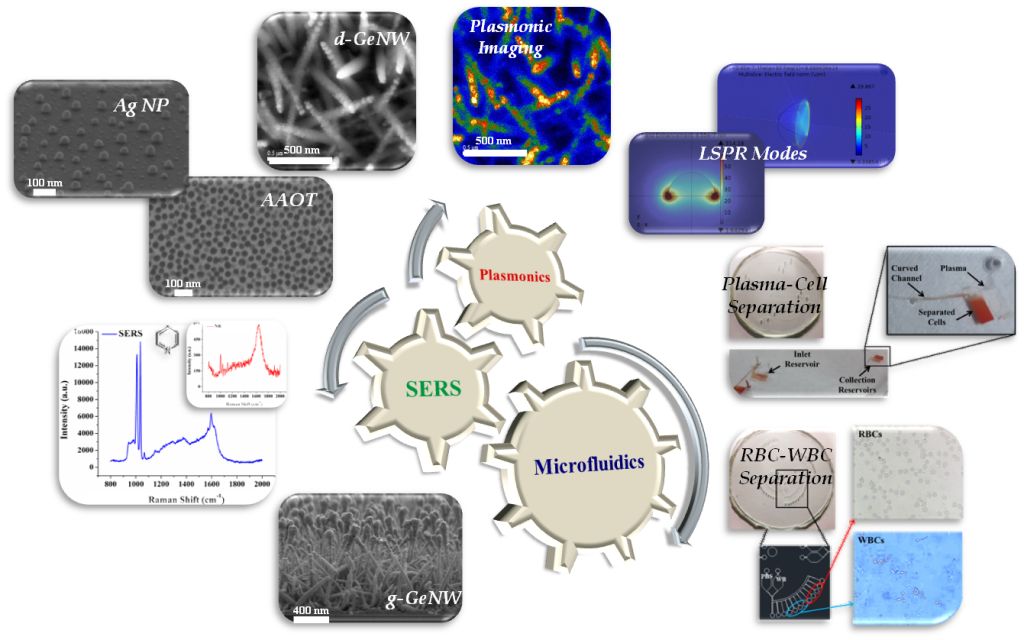
We are interested in investigating the localized surface plasmon resonance (LSPR) modes of metal nanostructures through simulation (COMSOL) and experiment (CL Imaging). Establishing effects of substrate permittivity has enabled us to engineer effective substrates with Ag nanoparticles (AgNP), Anodized Aluminium Oxide Templates (AAOT), AgNP deposited or grown on Germanium Nanowires (d-GeNW, g-GeNW) to name a few.
We are working on centrifugal microfluidics (LOACD) instead of conventional pressure driven microfluidics to avoid the disadvantages related to driving method (usage of expensive syringe pump) and footprint of the setup. We have developed centrifugal microfluidic devices for cell component separation.
We are in a process to integrate both the above mentioned tools and techniques to formulate device/s which can have the capability ranging from laboratory analysis to point-of-care testing. This work has been carried out with our collaborator, Prof. Navakanta Bhat at Centre of Nanoscience and Engineering (CeNSE).
Students:
Bio-photonics Lab
Study of biological systems combined with photonics
Light for Life

Bio-photonics lab serves a platform for interdisciplinary research where vibrational micro-spectroscopy (IR and Raman) is used to explore various aspects of biochemistry and medicine. We attempt to understand fundamental aspects of biophysical chemistry e.g., interaction of drugs on cancer cells, neuronal cells, bacteria etc. effect of SDS on hair; understanding the structure and dynamics of proteins and DNA. In terms of application based research we focus on sensitive and specific detection of various stages of cancer, sepsis; metabolic syndrome and identification of single bacteria with an aim towards detection of TB. We aspire to take vibrational micro-spectroscopic techniques to clinical bench; which would aid humanity and society for a better livelihood.
Students:
Terahertz Raman Spectroscopy
Witnessing Ultra Low Frequency Regime
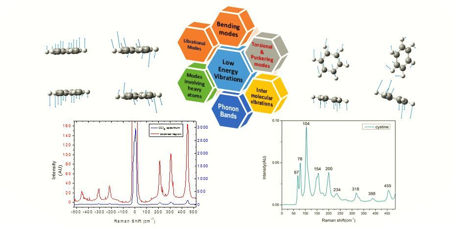
Molecular vibrations provide a straightforward approach to identify chemical signatures of various moieties present within a molecule. Because of practical difficulties, spectroscopists using IR absorption or Raman scattering are focusing mostly on fingerprint vibrations, which are highly selective for individual functional groups. However, there are several different motions which fall in the category of collective vibrations or reflect weak intermolecular interactions. These vibrations are extremely low in energy (0.1-10 THz or 3-300 cm-1) and it is very difficult to detect them with traditional instrumentation. We have developed a Terahertz Raman spectrometer based on Volume Bragg Gratings (VBGs) to detect these low-energy vibrational modes. The performance of the spectrometer was characterized using standard model compounds namely powdered sulphur and L-cystine.
Students:
Photochemistry of Hetero Aromatic Carbonyls
ns-Time-Resolved Absorption and Resonance Raman Spectroscopy
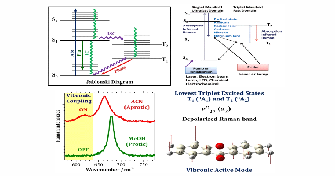
Photochemistry plays a key role in day to day life, like sunscreens which contain organic compounds that can absorb in UVA (400-315 nm) region of solar electromagnetic spectrum (not absorbed by ozone layer) and protect us from skin lesions. The potency of these sunscreens depends on their ability to absorb UV light without undergoing photodegradation (triplet states or long-lived radical species). When a molecule absorbs an UV photon, an electron from HOMO is promoted to LUMO of the molecule and leading to an electronically excited state. The fate of the excited electronic state will be governed by various photophysical and photochemical processes. There are various factors like substituent effect, solvent effect etc. that can govern these processes but our group is mainly focused on how a solvent can influence these processes.
Generally, for a bimolecular photochemical reaction to happen the reactants should encounter at a molecular scale and therefore diffusion of reactants is the bottleneck. In solution phase, the diffusion of molecules typically takes place in the nanosecond (ns) timescale and for a reaction to happen, the reactants have to undergo several collisions typically of 100-1000 s and therefore the excited state should be long-lived for example in microseconds (μs). Triplet states are long-lived typically of the order of several μs. In order to study the influence of solvent on the fate of these transient species, we perform ns-time-resolved spectroscopic techniques like time-resolved electronic absorption and molecular structure-specific resonance Raman spectroscopy.
Students:
UMARS Spectroscopy and its Applications
UMARS: Universal Multiple Angle Spectroscopy
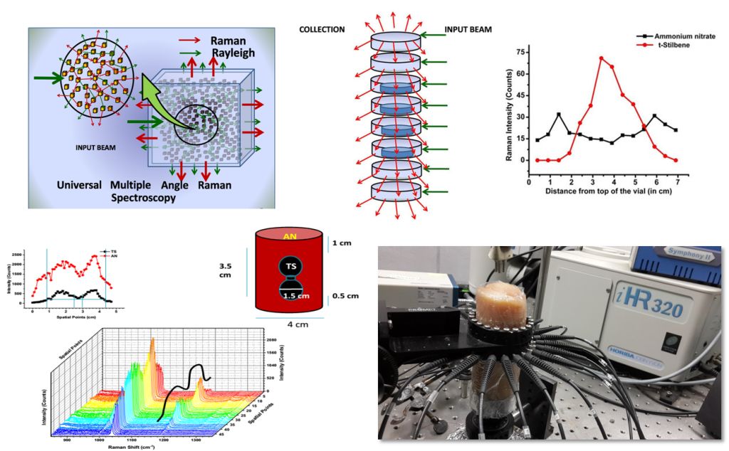
UMARS: Universal Multiple Angle Raman Spectroscopy is a technique in which collection of Raman signature from any direction irrespective of source position is possible. This works on multiple scattering principles, using many collection angles simultaneously to probe any type of scattering samples at varying depths using optical fibers. This technique allows Detection of Raman signals from all the observable angles and provides more depth of penetration which has potential applications in non invasive 3D imaging in materials and bio-medical optical imaging.
Our work mainly deals with 3D Raman imaging in biomedical and security applications. In
Biomedical applications we mainly deal with tumor detection, tissue imaging, and bone and skin disorder detection. We also focus on improvement of imaging at high depth in turbid medium. In Security applications we look for explosive and harmful chemical detection hidden inside a container.
Our lab is equipped with a laser system of 830, 785nm, a high resolution imaging spectrometer with three tuneable gratings, fiber optics for illumination and Raman signal collection, optics including lenses, mirrors, filters etc, optomechanics including fiber holders, translation and rotation stage mounts etc. In electronics we have oscilloscope, variable power supply, electronics components, FPGA boards etc.
Students: